Ingrid Kreissig, the story of a creative partnership that set a milestone in retinal surgery
VIEW DOCTOR PROFILE
Ingrid Kreissig
Ingrid Kreissig is a world-renowned retina specialist, currently Professor of Ophthalmology at the University of Mannheim-Heidelberg, Germany, Adjunct Professor of Clinical Ophthalmology at the Weill Cornell Medical College of New York, USA…

In this video, Prof. Ingrid Kreissig tells us the fascinating and many-year lasting story of a technique that subsequently revolutionized retinal detachment surgery. A story that shows how perseverance, a fascinating enthusiasm and rigorous scientific approach coupled with creative thinking, eventually lead to the success.
Minimal segmental buckling without drainage minimizes surgical trauma, causes no decompression of the eye, limits surgery to the area of the retinal break, harbors low morbidity and leads to fast recovery of vision. It is a low-budget technique does not require expensive surgical instruments or tamponades and is performed under local anesthesia.
Based on the principle of Ernst Custodis, introduced in 1963, this minimal extraocular technique, however, suddenly demonstrated serious postoperative complications. Therefore, it was first modified by Harvey Lincoff with the introduction of the sponge in 1965, and in the following years together with Ingrid Kreissig further modified and refined for its subsequent acceptance.
Though nowadays vitrectomy is a widely performed procedure for repair of retinal detachment, minimal segmental buckling without drainage or minimal extraocular surgery still remains the technique with the lowest rate of reoperation and morbidity. However, it requires a meticulous and time-consuming preoperative examination of the retina to limit the treatment to the area of the break and achieving postoperative reattachment. 1

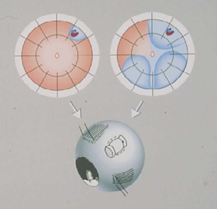
Fig. 1 – With minimal segmental buckling, a segmental elastic sponge is applied over the area of the retinal break(s). Thanks to the elasticity of the sponge buckle, no drainage of subretinal fluid is required. Coagulation therapy, either cryopexy or laser, is also limited to the area of the break(s). A variation of the technique entails the application on the break/s by a temporary balloon buckle which is not sutured onto the sclera and is withdrawn after a week 2. Image reproduced with the kind permission of Prof. Ingrid Kreissig.
1 Kreissig I. A practical guide to minimal surgery for retinal detachment. Vol 1: Diagnostics, segmental buckling without drainage, case presentations. Stuttgart-New York, Thieme, 2000: pg 1-288.
2 Kreissig I. A practical guide to minimal surgery for retinal detachment. Vol. 2: Temporary tamponades with balloon and gases without drainage, buckling versus gases versus vitrectomy, reoperation, case presentations. Stuttgart-New York, Thieme, 2000: pg 1-356.